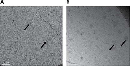
Figure 5.

Cryo-TEM images of (A) BSA-HA conjugates (40 μM) without PTX. The grainy surface indicates BSA-HA conjugates (small arrows) and larger gray stains indicate self-assembled aggregates of BSA-HA conjugates (large arrows). (B) 2:1 molar ratio of PTX (80 μM) with BSA-HA conjugates; All samples contained 0.8% DMSO in PBS. The molar ratio of PTX and BSA-HA conjugates was 2:1. PTX aggregates and nanocrystals were coated with BSA-HA conjugates adsorbed to their surfaces (arrows in Figure 5B).
