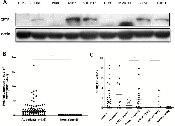
Figure 1. CFTR expression in leukemia cell lines and primary leukemia samples.

(A) CFTR protein expression in leukemia cell lines was detected by Western blotting with HBE as a positive control (value = 1) and HEK293 as a negative control. (B) CFTR protein expression in primary leukemia samples (n = 138) and normal human MNCs (n = 50) was detected by Western blotting. Significance compared with the MNC group is indicated. Data are given as the mean ± SEM relative to HBE expression. ***P < 0.001. (C) Among 40 cases of B-ALL and 12 cases of CML primary leukemia cells, the expression of CFTR in 20 cases of Ph+ B-ALL and 6 cases of CML-BC was higher than that in 20 cases of Ph- B-ALL and 6 cases of CML-CP. *P < 0.05.
