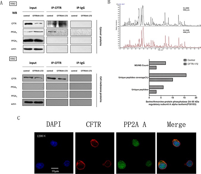
Figure 5. The interaction between CFTR and PP2AA subunits was interrupted by CFTRinh-172.

(A) K562 cells were exposed to CFTRinh-172 (150 μM, 24 h) and were lysed to extract the cytosolic and membrane-associated proteins. PP2A subunits were immunoprecipitated with CFTR and detected by Western-blot assay using monoclonal antibodies specific to the CFTR, PP2AA subunit and PP2AC subunits. (B) LC-MS analysis confirmed the presence of the PP2AA subunit in IP-CFTR products between 50 kd and 70 kd and revealed a decrease in PP2AA subunit in IP-CFTR products after treatment with CFTRinh-172. The column graph shows the signal attributed to the PP2AA subunit(PR65) in two report lists. Top: the control group; bottom: the CFTRinh-172 treatment group. (C) CFTR and PP2AA subunit exhibited marked co-localization in the cytosol as detected by IF co-localization analysis. K562 cells were stained with mAbs against CFTR (red) and the PP2AA subunit (green) as well as DAPI(blue). Fluorescence images were collected using a confocal microscope. Orange indicates the colocalization of CFTR and PP2AA(magnification, ×1200).
