Figure 1.

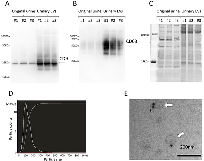
A, B. Western blotting showed the expression of specific proteins ((A) CD9 and (B) CD63) in urinary EVs. C. A representative gel stained with SYPRO Ruby, showing the protein profile of original urine and urinary EVs. D. Nanoparticle tracking analysis revealed that almost all of the particles extracted by ultracentrifugation were under 200 nm in size. E. Electron microscopy shows urinary EVs immunolabeled with anti-CD9 antibody conjugated by 20-nm protein gold nanoparticles (white arrows indicate EVs, and the asterisk indicates gold nanoparticles). Data are representative of 3 (A, B, C and D) and one (E) independent experiment.
