
Figure 3. Pediatric brain tumor cells treated with carmofur underwent apoptosis as demonstrated by the Annexin-V-Alexa-488 conjugate staining.
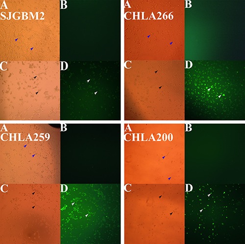
Microscopy studies pediatric brain tumors are shown. Top left panel, SJGBM2; top right panel, CHLA266; bottom left panel, CHLA259; bottom right panel, CHLA200; control cells imaged with the brightlight (A), with the fluorescent light (B) vs cells treated (12 hrs) with 50 μM carmofur imaging with the brightlight (C), with the fluorescent light (D). Brightlight imaging of control live cells (blue arrows) and death cells (black arrows) are shown. Whereas a large number of apoptotic cells stained with Annexin-V-Alexa-488 (white arrows) were observed under fluorescent imaging when treated with carmofur, very little to no staining was seen in control untreated cells (B).