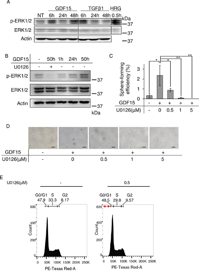
Figure 3. Sustained activation of ERK1/2 appears to be required for GDF15-induced tumor sphere formation.

A. Immunoblotting analysis of phosphorylated ERK1/2 (p-ERK1/2) and ERK1/2 expression in MCF7 cells treated with GDF15 (200 ng/mL) or TGFβ1 (200 ng/mL). NT, not treated. Actin was used as a loading control. The lysate of MCF7 cells stimulated with heregulin (HRG) was used as a positive control. B. Immunoblotting analysis of p-ERK1/2 and ERK1/2 expression in MCF7 cells treated with GDF15 (200 ng/mL) in the presence or absence of U0126 (5 μM). C. Sphere formation assay of GDF15-treated (200 ng/mL) MCF7 cells in the presence or absence of the indicated concentration of U0126. n=4. **P < 0.01, *P < 0.05. D. Representative images of (C). Scale bar: 100 μm. E. Cell cycle analysis of MCF7 cells treated with GDF15 (200 ng/mL) in the presence or absence of U0126 (0.5 μM). Apoptotic cells are observed in the region indicated with the red arrows (the sub-G1 area).
