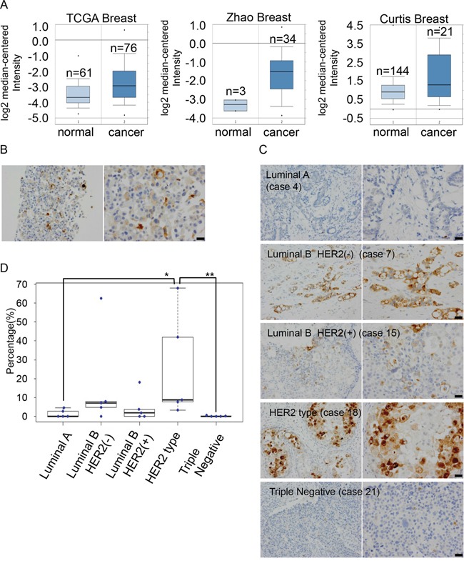
Figure 5. Expression levels of GDF15 are heterogeneous among cancer cells in MCF7 cells and human breast cancer tissues.

A. Analysis of the expression levels of GDF15 transcripts in Oncomine database. B. Immunohistochemical staining of GDF15 in MCF7 cells in paraffin blocks using an anti-GDF15 antibody. Left, original magnification 200x. Right, scale bar: 20 μm. C. Immunohistochemical staining of GDF15 in various subtypes of breast cancer tissues. Left, original magnification 400x. Right, scale bar: 20 μm. D. Box plots of GDF15 expression among 25 clinical breast cancer tissues that include 5 cases in each subtype. **P < 0.01, *P < 0.05.
