
Figure 5. IMQ combined with IR enhances lymphocyte infiltration but decreases MDSC abundance in tumor lesions.
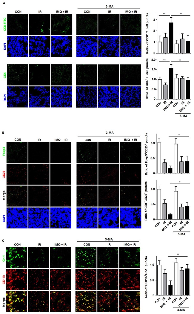
Immunostaining was performed for each tumor section 21 days after B16F10 cell inoculation. A. Infiltrated T cells were observed in the frozen tumor sections. The tissues were stained with DAPI to visualize nuclei (blue) and were immunolabeled with anti-CD4 and anti-CD8 antibodies, which were detected via the addition of FITC-conjugated IgG (green). The numbers of CD8+ and CD4+ T cell puncta were determined under a confocal microscope and are presented as proportions relative to the number of corresponding cells in the control group. B. The Treg population was detected with anti-CD25 (PE-conjugated, red) and anti-Foxp3 (FITC-conjugated, green) antibodies. The Treg numbers were counted in merged images captured via confocal microscopy. C. The MDSC population was detected using anti-CD11b (PE-conjugated, red) and anti-Gr1 (FITC-conjugated, green) antibodies. For all the experiments, the mean values ± SE from eight measurements are shown. Significant differences are indicated by *p < 0.05, **p < 0.01, and ***p < 0.001.