
Figure 1. Tan I exerts cytotoxicity and induces autophagy in H28 and H2452 mesothelioma cells.
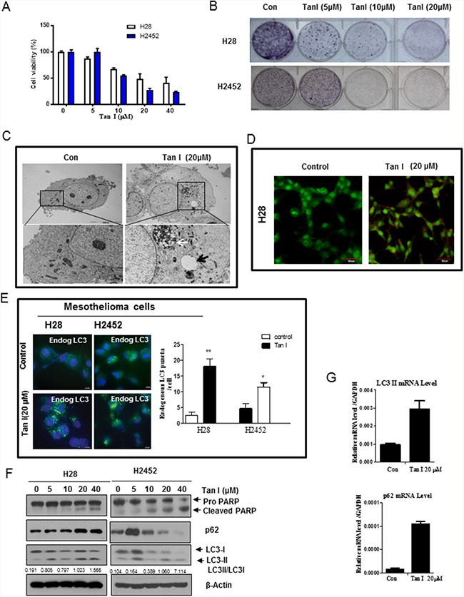
A. Cytotoxicity of Tan I in the H28 and H2452 mesothelioma cell lines. Two mesothelioma cell lines were treated with various concentrations of Tan I (0, 5, 10, 20, and 40 μM) for 24 h. Cell viability was determined by the MTT assay. B. The inhibitory effect of Tan I on colony formation in H28, and H2452 mesothelioma cells. The colony formation assay was performed on mesothelioma cells treated with Tan I (20 μM). C. Effect of Tan I on autophagic vacuoles in H28 cells by TEM observation. H28 cells were treated with Tan I (20 μM) for 24 h, and autophagic morphology was observed by TEM. The black arrow indicates an autolysosome that contains the remnants of digested organelles. High-magnification image of the black square (bottom panel). D. Effect of Tan I on acidic autophagic vacuoles in Tan I-treated H28 cells. H28 cells were treated with Tan I for 24 h, stained with acridine orange (AO) and observed under the FLUOVIEW FV10i confocal microscopy (Olympus, Tokyo, Japan). E. Effect of Tan I (20 μM) on punctae formation of endogenous LC3II in H28 and H2452 cells by immunofluorescence. Bar scale = 10 μM, DAPI-blue, endogenous LC3-Green. F. Effect of Tan I on PARP, p62 and LC3II in H28 and H2452 mesothelioma cells. G. Effect of Tan I on the mRNA expression of p62 and LC3II in H28 cells by RT-qPCR. Total RNA was isolated from Tan 1 treated H28 cells and RT-PCR was performed as shown in Materials and Methods.