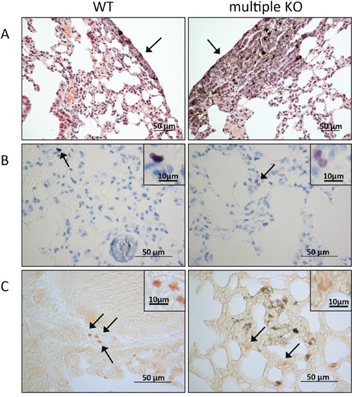
Figure 2. Histological analysis of lung tissue.

On day 14 post B16F10 cell injection, lungs were paraffin imbedded, sectioned and stained with hematoxylin and eosin A. toluidine blue B. or for chloroacetate esterase activity C. (A) Arrows point to tumor areas. Note a larger tumor area in the section from multiple KO mice (right) in comparison with WT controls (left); original magnification x 200; bar = 50 μm. (B) Toluidine blue-positive MCs (marked by arrows) are present in lung sections from both WT (left) and multiple KO (right). Inserts show a higher magnification of the toluidine blue-stained MCs; note that MCs in lungs of multiple KO mice stain less intensely in comparison with WT MCs. (C) Chloroacetate esterase staining shows the presence of chymase-positive MCs in lung of both WT (left) and multiple KO (right) mice (marked by arrows): insets show larger magnifications of chloroacetate esterase-stained MCs; note that MCs in lungs from multiple KO mice stain less intensely with chloroacetate esterase, indicating lower content of chymase activity. Original magnification x 400; bars = 50 μm; bars in the inserts =10 μm.
