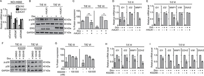
Figure 5. ALK1 inhibitors decrease T/E-induced ALK1 signaling.

(A) ERG knockdown in NCI-H660 cells (50 nM) showed reduced levels of TGFB1 and ALK1. (B) Western blot analysis in LNCaP-T/E cells revealed reduced p38 phosphorylation after treatment with rhALK1 (5μg/mL) compared to PBS-treated control cells. (C) p-p38/p38 ratios after densitometric analysis of Western blot bands shown in (B) in T/E III and T/E VI cells, respectively. (D–E) Expression levels of TGF-β-responsive genes in (D) T/E III and (E) T/E VI expressing cells after rhALK1 treatment (5μg/mL) determined by qPCR. (F) Western blot analysis of p38 phosphorylation after treatment with K02288 (at indicated concentrations) compared to DMSO-treated control cells. (G) p-p38/p38 ratios after densitometric analysis of Western blot bands shown in (F) of T/E III and T/E VI cells, respectively. (H–I) Expression levels of TGF-β/BMP-responsive genes in (H) T/E III and (I) T/E VI expressing cells after simultaneous treatment with Dox and K02288 (500nM) were determined by qPCR.
