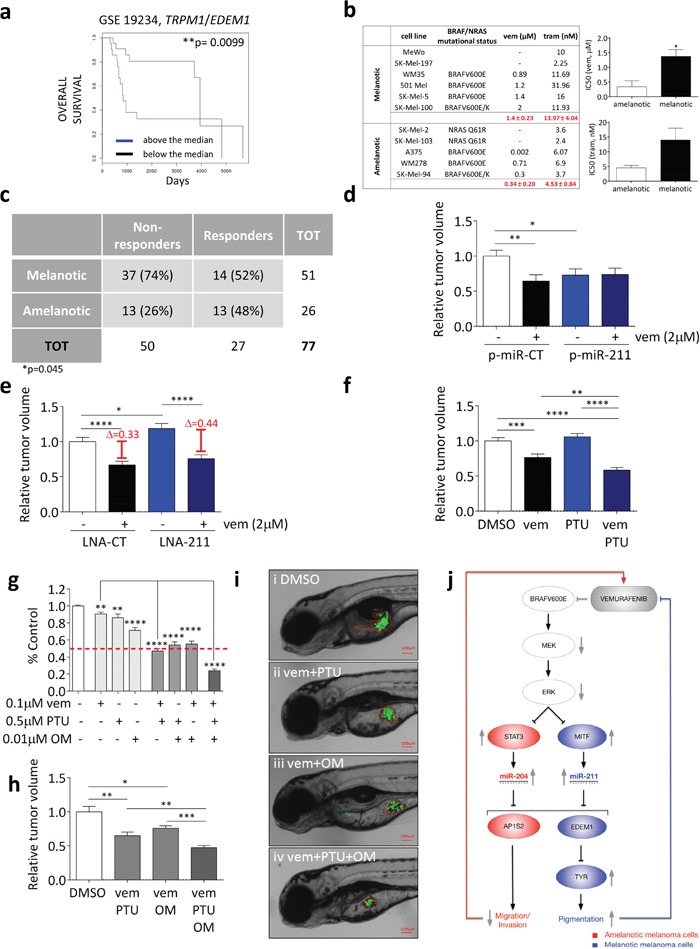
Figure 9. Pigmentation impairs the activity of BRAFi and MEKi.

(a) Above the median (blue) and below the median (black) TRPM1/EDEM1 ratios allow to stratify metastatic melanoma patients according to their overall survival (GSE 19234). (b) IC50 of melanotic and amelanotic melanoma cell lines treated with vemurafenib (BRAFV600 E or K) and trametinib (BRAFV600 E or K, wt BRAF). (c) Response of 51 metastatic melanoma patients with melanotic tumors and 26 metastatic melanoma patients with amelanotic tumors to treatment with BRAFi or combined BRAFi/MEKi. Patients showing disease progression upon treatment were classified as non-responders, while patients showing stable disease, partial response or complete response upon treatment were classified as responders. The Fisher Exact Probability Test indicate that patients with melanotic metastases are significantly more likely to be non-responders to targeted therapy compared to patients with amelanotic metastases (p=0.045) (d) 501 Mel cells that stably over-express a control miRNA (p-miR-CT, white and black) or miR-211 (p-miR-211, blue and dark blue) were treated with DMSO or 2uM vemurafenib for 48h. They were then injected into the yolk sac of 48hfp zebrafish embryos. The masses of the xenografted tumors were measured 48h later. (e) 501 Mel cells transiently transfected with LNA-CT (white and black), or LNA-211 (blue and dark blue) were treated with DMSO or 2uM vemurafenib for 48h. They were then injected into the yolk sac of 48hpf zebrafish embryos. The masses of the xenografted tumors were measured 48h later. (f) 501 Mel cells were treated with DMSO (white), 0.2uM vemurafenib (black), 0.1uM PTU (blue) or 0.2uM vemurafenib plus 0.1uM PTU (dark blue) for 48h. They were then injected into the yolk sac of 48hpf stage zebrafish embryos. The masses of the xenografted tumors were measured 48h later. (g) Cell number upon the treatment of 501 Mel cells with 0.1uM vemurafenib, 0.5uM PTU and 0.01uM oligomycin (OM) or their combinations for one week. (h-i) 501 Mel cells were treated with DMSO (white), 0.1uM vemurafenib plus 0.5uM PTU, 0.1uM vemurafenib plus 0.01uM oligomycine or 0.1uM vemurafenib plus 0.5uM PTU plus 0.01uM oligomycine (dark grey) for 48h. They were then injected into the yolk sac of 48hpf zebrafish embryos. (h) The masses of the xenografted tumors were measured 48h later. (i) Representative pictures of the tumor masses. (j) Cartoon that summarizes the main findings of this article. miR-204 and miR-211 are negatively regulated by BRAFV600E through the ERK pathway and are under the transcriptional control of STAT3 and MITF, respectively. By targeting AP1S2, miR-204 is the mediator of the anti-motility activity exerted by vemurafenib on amelanotic cells. Conversely, by targeting EDEM1 and hence preventing TYR degradation through the ERAD pathway, miR-211 is the mediator of the pro-pigmentation activity exerted by vemurafenib on melanotic cells. Such an activity in turn limits the efficacy of vemurafenib itself. The graphs represent the mean±SEM of 3 independent experiments. *p<0.05, **p<0.01, ***<0.001, ***<0.001.
