Fig. 4.
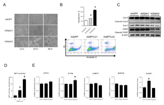
Adenoviral overexpression of SPTLC2 induces apoptosis in HepG2 cells but does not activate ER stress. HepG2 cells were infected with SPTLC1 or SPTLC2 adenoviruses at 2 MOI for 24 and 48 hours. Cell morphology was changed only after infection of SPTCL2 adenovirus (A). After 24 hours of infection, cells were stained with Annexin V and propidium iodide and the degree of apoptosis quantified by flow cytometry (B). Data are presented as the mean ± SEM. n = 3. *P < 0.05. HepG2 cells were infected with adenoviruses at 2 MOI for 24 hours and whole-cell lysates were subjected to immunoblotting analyses of caspase-3 (Cas 3), caspase-9 (Cas 9), and PARP (C). β-actin was used as a control. Under the same condition, SPT enzyme activity was measured as described in Materials and Methods (D). Then, the expression of ER stress genes including ATF4, ATF6, sXBP1, GRP78, and CHOP was measured by quantitative real-time PCR (E). Amount of mRNA was normalized by β-actin. Data are presented as the mean ± SEM. n = 3. *P < 0.05.

