Figure 1.
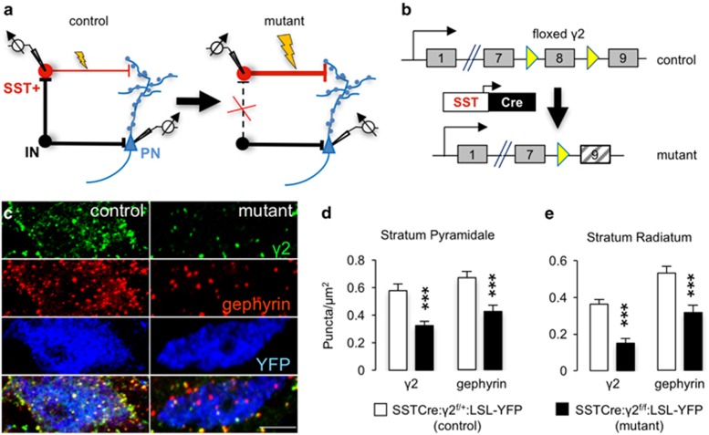
Deletion of postsynaptic γ-aminobutyric acid type A receptors (GABAARs) and gephyrin from somatostatin-positive (SST+) neurons of SSTCre:γ2f/f mice. (a) Strategy for γ2 subunit knockout-mediated disinhibition of SST+ interneurons. Loss of synaptic GABAARs removes inhibitory input (IN) to SST+ neurons and increases excitability of these neurons. Increased excitability of SST+ neurons strengthens inhibitory synaptic inputs to apical dendrites and spines of pyramidal cells (PN). (b) Schematic of Cre-mediated inactivation of the ‘floxed' γ2 locus. (c) Representative micrographs of the soma of an SST+ neuron from a SSTCre: γ2f/+:LSL-YFP control mouse (left column) compared with a SST+ neuron from a SSTCre:γ2f/f:LSL-YFP mutant animal, immunostained for the γ2 subunit (top row, green), gephyrin (second row; red) and yellow fluorescent protein (YFP; third row; blue) with merged images showing colocalization of γ2 and gephyrin in yellow in the bottom row. Note the drastic reduction in punctate staining for both the γ2 subunit and gephyrin, indicative of loss of functional synapses. Residual staining for γ2 is likely attributable to dendrites of Cre-lacking neighboring neurons. (d) Quantification of puncta densities overlapping with YFP+ cell somata (puncta per μm2) in S. pyramidale and S. radiale of the hippocampus. Densities for both proteins were significantly reduced in both areas (P<0.001, respectively). ***P<0.001, Mann–Whitney, n=30–40 cells, 2 mice/genotype.

