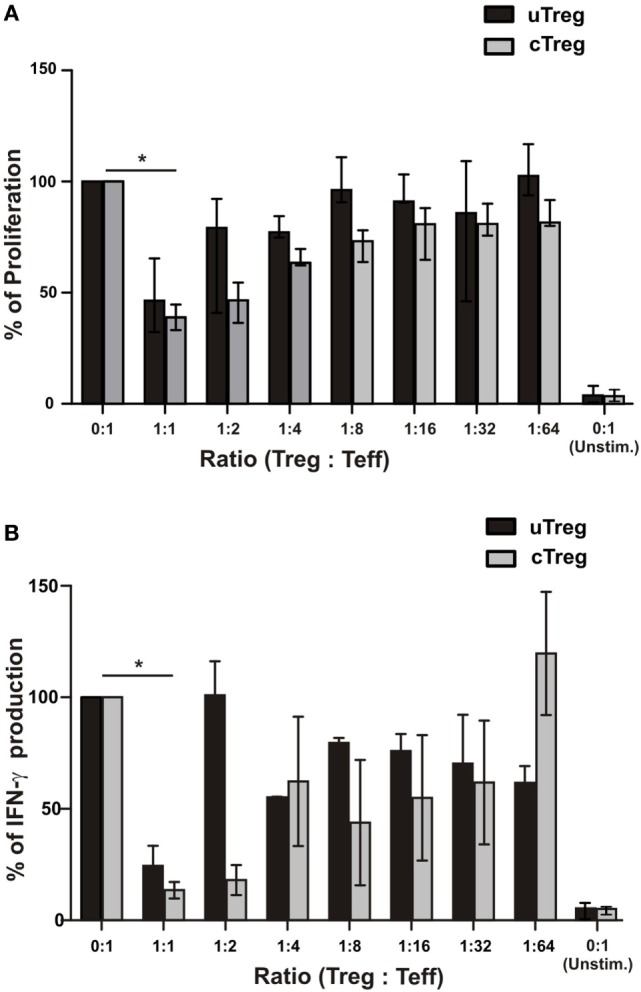
Figure 7.

Functional characterization of uTregs. FACS-isolated CD4+CD25−CD39− effector T cells were stimulated with anti-CD3 plus allogeneic mitomycin-treated PBMCs and cocultured with CD4+CD25−CD39+ uTregs or CD4+CD25+CD39+ cTregs cells at the indicated Treg:Teff ratios. In addition, effector T cells were left unstimulated in order to determine basal proliferation. After 5 days, proliferation and IFN-γ secretion were evaluated. Proliferation levels of stimulated CD4+CD25−CD39− effector T cells without any Treg population were defined as 100%. (A) Proliferation of effector T cells at different Teff/Treg ratios. (B) IFN-γ production in cell-free supernatants from cocultures was assessed by ELISA. Bars represent mean values + SEM from three individual experiments. *p < 0.05.
