Fig. 6.

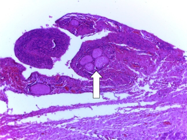
A photomicrograph of the ranula specimen following H&E low power (×10) showing extracellular pools of salivary mucin (arrow) surrounded by inflammatory cells and fibrosis

A photomicrograph of the ranula specimen following H&E low power (×10) showing extracellular pools of salivary mucin (arrow) surrounded by inflammatory cells and fibrosis