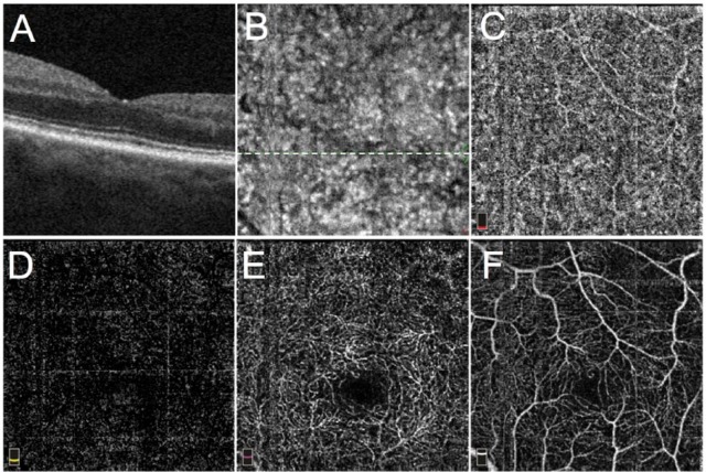
Figure 1.
Optical coherence tomographic (OCT) angiography in a healthy right eye of a 74-year-old man: (A) spectral domain OCT B scan through the fovea (corresponding to the white dotted line in B), (B) en face structural OCT, (C) 3 × 3 mm spectral domain OCT angiogram of the choriocapillaris centered on the fovea (acquired using RTVue XR Avanti; Optovue Inc., Fremont, CA, USA) with split-spectrum amplitude-decorrelation angiography software, (D) OCT angiogram of the outer retina showing absence of blood flow, (E) OCT angiogram of the deep capillary plexus, and (F) OCT angiogram of the superficial capillary plexus.

