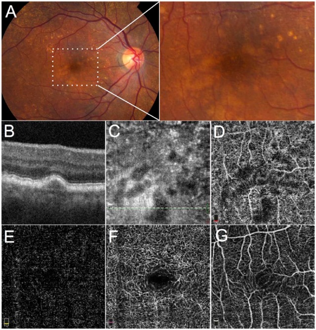
Figure 2.
Optical coherence tomographic (OCT) angiography in nonexudative age-related macular degeneration in the right eye of an 80-year-old woman, with exudative AMD in the fellow eye (not shown). (A) Color fundus photograph showing numerous drusen, enlarged to approximate area of following OCT angiograms; (B) SD-OCT B scan through a drusen (corresponding to the white dotted line in B); (C) en face structural OCT showing dark areas corresponding to drusen; (D) 3 × 3 mm spectral domain optical coherence tomographic (SD-OCT) angiogram of the choriocapillaris showing shadowing artifact and/or flow impairment in areas under the drusen; (E) OCT angiogram of the outer retina showing absence of choroidal neovascularization; (F) OCT angiogram of the deep capillary plexus appears uninvolved; and (G) OCT angiogram of the superficial capillary plexus appears uninvolved.

