
Figure 4.
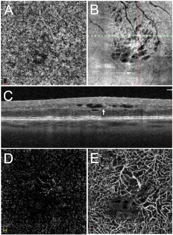
Type III choroidal neovascularization (CNV) in exudative age-related macular degeneration (retinal angiomatous proliferation) in the left eye of a 68-year-old man, treatment-naive. (A) 3 × 3 mm spectral domain optical coherence tomographic (SD-OCT) angiogram of the choriocapillaris showing patchy loss of flow signal due to overlying intraretinal fluid; (B) en face structural optical coherence tomography (OCT) of the same area showing patchy hyporeflectivity corresponding to intraretinal cystic fluid, as well as hyperreflectivity corresponding to retinal blood vessels; (C) OCT B scan through area of intraretinal fluid and type III CNV (arrow) (corresponding to the white dotted line in B). Width of OCT B scan is equivalent to width of angiograms in (A), (D), and (E). (D) OCT angiogram of the outer retina shows part of the CNV centrally. (E) OCT angiogram of the deep capillary plexus showing dilated CNV vessel with surrounding loss of flow signal due to intraretinal fluid.