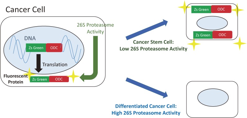
Figure 3.
Cancer stem cells can be visualised because they have downregulated 26S proteasome activity. Cells are transfected with a vector coding for a fusion protein consisting of ZsGreen, and the C-terminal degron of the ornithine decarboxylase. Degron directs the destruction of the fluorescent protein by proteasomes in differentiated cancer cells. In cancer stem cells, the fusion protein is not destroyed and the cells are fluorescently labelled.

