Figure 1.

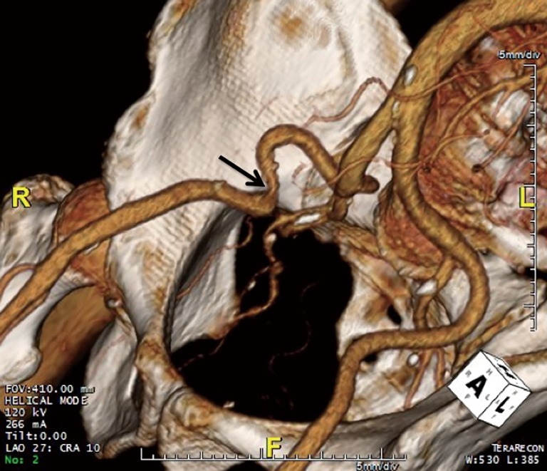
Three-dimensional (3D) volume rendered image obtained from a computed tomography (CT) scan showing a tortuous right external iliac artery with a narrowing (indicated by the arrow). In contrast, the left external iliac artery is normal.

Three-dimensional (3D) volume rendered image obtained from a computed tomography (CT) scan showing a tortuous right external iliac artery with a narrowing (indicated by the arrow). In contrast, the left external iliac artery is normal.