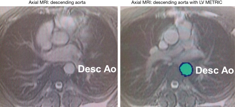
Figure 1.
Assessment of aortic physiology via partial voxel automated segmentation. Axial cine-MRI without automation (A) and with automation (B) in the mid-descending aorta. Partial voxel interpolation (PVI) of the aortic segment is assessed at both the largest (systole) and smallest (diastole) area size.

