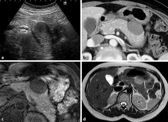
Fig. 1.

a Abdominal ultrasound shows a well-defined hypoechoic mass measuring 30 mm in diameter with internal heterogeneity in the pancreatic tail. b Abdominal contrast-enhanced CT (early phase) shows a well-defined mass lesion measuring approximately 30 mm in diameter, extending from the gastric wall to the pancreatic tail. c, d Abdominal MRI shows a mass lesion with low signal intensity on T1-weighted imaging (c) and slightly high signal intensity on T2-weighted imaging (d).
