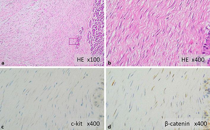
Fig. 3.

a, b Histopathological findings show cord-like proliferation of spindle-shaped cells with minimal atypia, and the intercellular spaces are filled with thick bundles of collagen fibers. a HE stain, ×100. b HE stain, ×400. Immunostaining shows that the cells are negative for c-kit (c) and positive for nuclear β-catenin (d). c c-kit, ×400. d β-catenin, ×400.
