
Fig. 2.
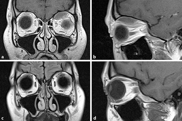
Coronal (a) and sagittal (b) T1-weighted MRI identifying a biconvex lesion in the superior medial orbital roof with compression of superior muscles. Frontal (c) and sagittal (d) T1-weighted FLAIR views 4 months later with complete resolution of the lesion.