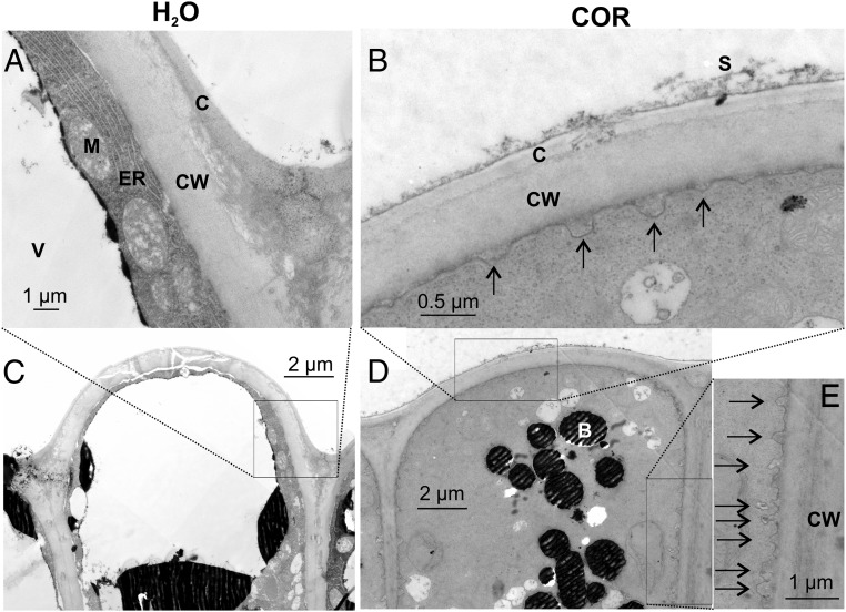
Fig. 1.
Exocytotic vesicle fusion is stimulated in activated gland complexes. EMs of the outer layer of resting (A and C) and COR-stimulated (B, D, and E) Dionaea gland complexes are shown. A detailed view (A, B, and E) and overview (C and D) are shown. Whereas resting glands only exhibit a few exocytotic events, a massive rise in exocytotic vesicle fusion with the plasma membrane (black arrows) could be detected 48 h after COR stimulation. B, dark-stained body; C, cuticle; CW, cell wall; ER, endoplasmic reticulum; M, mitochondria; S, secreted fluid; V, vacuole. Slight shadow lines are due to carrier film handling during TEM sample preparation; all images are noncomposite originals.

