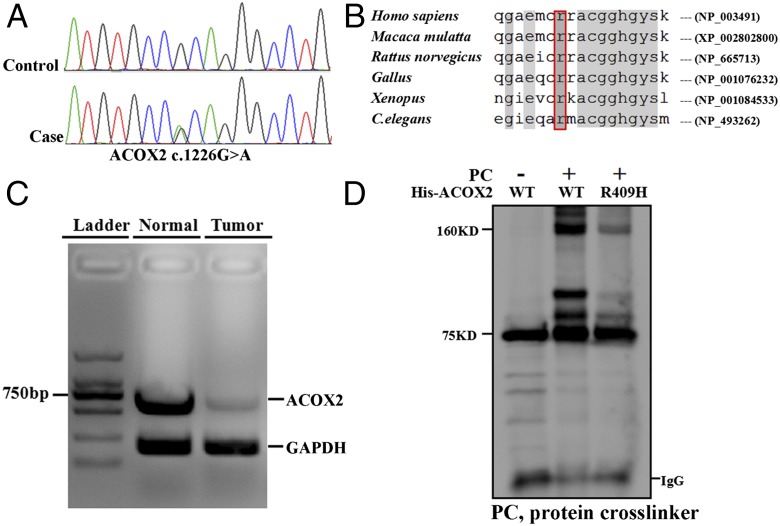
Fig. 1.
ACOX2 deficiency in a patient with a primary malignant cardiac tumor. (A) Sanger sequencing of ACOX2 (c.1226G > A, p.Arg409His) using blood samples from patient 1. (B) Sequence alignment of ACOX2 p.Arg409His (National Center for Biotechnology Information reference sequences are indicated). C. elegans, Caenorhabditis elegans. (C) ACOX2 and GAPDH were amplified from the cDNA of adjacent normal and tumor tissue from patient 1 as indicated. GAPDH served as a positive control to demonstrate the reduction of ACOX2 expression in tumor tissues. (D) ACOX2 (R409H) affected normal dimer formation. HEK293T cells were transfected with His-tagged wild-type or (R409H) ACOX2 for recombinant overexpression. A 1:100 dilution of a protein cross-linker (PC) was added to the RIPA buffer before cell lysis. Subsequently, anti-His immunoprecipitation and anti-ACOX2 immunoblotting were performed following standard procedures. A 75-KDa band was indicative of ACOX2-His recombinant protein. After PC treatment, a 160-KDa band was generated, corresponding to an ACOX2 dimer. This band was significantly decreased in cells transfected with mutant ACOX2.

