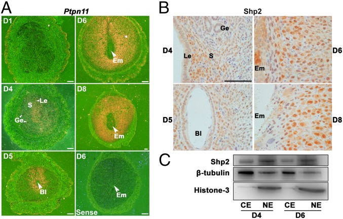
Fig. 1.
Shp2 is spatiotemporally expressed in periimplantation mouse uteri exhibiting a nuclear localization. (A) In situ hybridization of Ptpn11 mRNA in D1–4 uteri and D5–8 implantation sites. Shp2 mRNA is visible as the brown color. The white arrowhead indicates the embryo. (White scale bars, 250 μm.) (B) Immunohistochemical staining of Shp2 protein in D4 uteri and D5–8 implantation sites. The brown color indicates positive staining for Shp2. Note the dominant nuclear location of Shp2. (Black scale bar, 100 μm.) (C) Immunoblotting analysis of Shp2 protein in different cellular fractions from the D4 uteri and D6 implantation sites. β-Tubulin serves as a cytoplasmic loading control, whereas histone 3 serves as a nuclear loading control. Bl, blastocyst; CE, cytoplasmic extraction; Em, embryo; Ge, glandular epithelium; Le, luminal epithelium; NE, nuclear extraction; S, stroma. Data shown are representative of at least three independent experiments.

