Fig. 2.
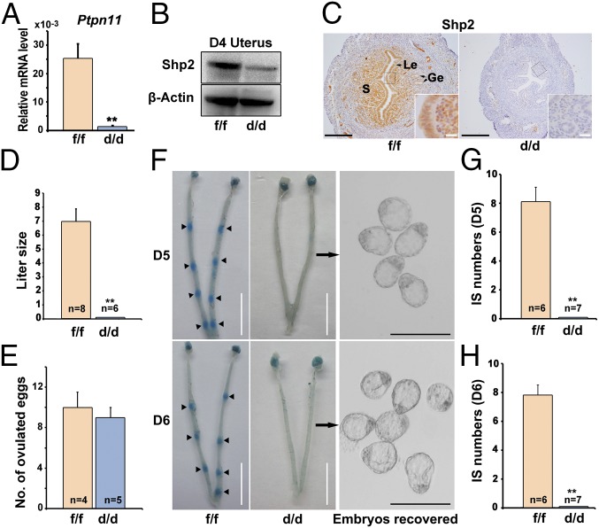
Uterine Shp2 deficiency induces complete implantation failure resulting in female infertility. (A) Real-time PCR analysis of Shp2 mRNA levels in D4 uteri from wild-type (Shp2f/f) and knockout (Shp2d/d) mice. The values are normalized to the Gapdh expression level and indicated as the mean ± SEM, n = 3. **P < 0.01. (B) Immunoblotting analysis of Shp2 protein levels in D4 Shp2f/f and Shp2d/d uteri. β-Actin is used as loading control. (C) Immunohistochemistry analysis of Shp2 protein in D4 Shp2f/f and Shp2d/d uteri. Ge, glandular epithelium; Le, luminal epithelium; S, stroma. (White scale bar, 25 μm; black scale bar, 250 μm.) (D) Average litter sizes in Shp2f/f and Shp2d/d female mice. Number within the bar indicated the number of mice tested. **P < 0.01. (E) Number of ovulated oocytes in Shp2f/f and Shp2d/d mice. Number within the bar indicates the number of mice tested. (F) Implantation status of Shp2f/f and Shp2d/d mice on D5–6. Implantation sites are visualized by blue dye method and the unimplanted embryos are recovered from Shp2d/d uteri (Right image). Black arrowheads indicate the implantation sites. Black arrows indicate the embryos in the right panels are recovered from the corresponding uteri. (White scale bar, 1 cm; black scale bar, 250 μm.) (G and H) Average number of implantation sites in Shp2f/f and Shp2d/d mice on D5 (G) and D6 (H) of pregnancy. Number within the bar indicates the number of mice tested. Mice that failed to recover any embryos are excluded in statistical analysis. IS, implantation site; **P < 0.01. Data shown are representative of at least four independent experiments.

