Fig. 4.
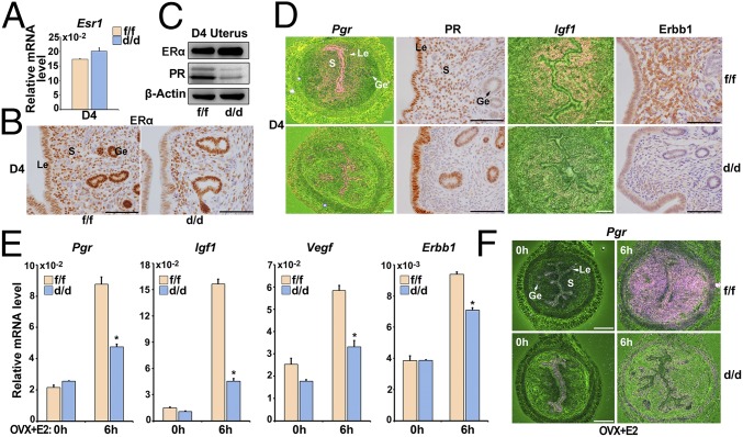
Loss of Shp2 compromises uterine estrogen responsiveness and thus PR expression. (A) Real-time PCR detection of the mRNA levels of Esr1 in D4 Shp2f/f and Shp2d/d uteri. The values are normalized to the Gapdh expression level and indicated as the mean ± SEM, n = 3. (B) Immunohistochemical staining of ERα in D4 Shp2f/f and Shp2d/d uteri. The brown color indicates the positive signal. (C) Immunoblotting analysis of ERα and PR in D4 Shp2f/f and Shp2d/d uteri. β-Actin is used as loading control. (D) In situ hybridization and immunohistochemical analysis of estrogen–ERα target genes in D4 Shp2f/f and Shp2d/d uteri. The brown color indicates the positive signal. (E) Real-time PCR detection of estrogen–ERα target gene expression levels in Shp2f/f and Shp2d/d ovariectomized mouse uteri in response to E2 treatment. The values are normalized to the Gapdh expression level and indicated as the mean ± SEM, n = 3. *P < 0.05. (F) In situ hybridization of Pgr in Shp2f/f and Shp2d/d ovariectomized mouse uteri after E2 treatment. The brown color indicates the positive signal. Ge, glandular epithelium; Le, luminal epithelium; S, stroma. (White scale bar, 250 μm; black scale bar, 100 μm.) Data shown are representative of at least three independent experiments.

