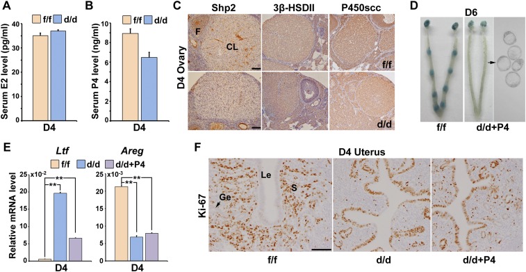
Fig. S1.
Progesterone supplementation failed to restore embryo implantation in Shp2d/d mice. (A) Serum estradiol-17β (E2) level in Shp2f/f and Shp2d/d mice on day 4 (D4) of pregnancy. (B) Serum progesterone (P4) level in Shp2f/f and Shp2d/d mice on D4 of pregnancy. Data are means ± SEM (*P < 0.05; Student’s t test). Numbers within bars indicate numbers of mice examined. (C) Immunohistochemistry staining of 3β-hydroxysteroid dehydrogenase II (3β-HSDII) and P450 cholesterol side-chain cleavage enzyme (P450scc) in Shp2f/f and Shp2d/d D4 ovaries. CL, corpus luteum; F, follicle. (Scale bars, 100 μm.) (D) Embryo implantation status of Shp2d/d mice as revealed by blue dye method on D6 of pregnancy after receiving daily P4 supplementation (2 mg per mouse) from D3–5. Black arrow indicates the embryos in the right panel are recovered from the corresponding uteri. (E) Real-time PCR analysis of the uterine receptivity marker genes on D4 after P4 supplementation. The values are normalized to the Gapdh expression level and indicated as the mean ± SEM, n = 3. **P < 0.01. (F) Immunohistochemical staining of Ki-67 in Shp2f/f and Shp2d/d in D4 uteri. (Scale bar, 100 μm.) Ge, glandular epithelium; Le, luminal epithelium; S, stroma.

