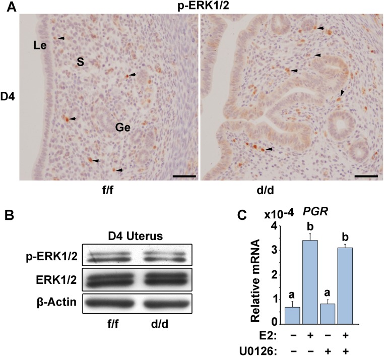
Fig. S3.
ERK1/2 expression and phosphorylation remain at comparable levels in D4 Shp2f/f (f/f) and Shp2d/d (d/d) uteri. (A) Immunostaining of phospho-ERK1/2 (p-ERK1/2) in Shp2f/f and Shp2d/d uteri on D4 of pregnancy. (Scale bars, 100 μm.) Black arrowheads indicate the positive cells. Ge, glandular epithelium; Le, luminal epithelium; S, stroma. (B) Immunoblotting analysis of p-ERK1/2 and total ERK1/2 expression in Shp2f/f and Shp2d/d uteri on D4 uteri. β-Actin serves as a control. (C) Real-time PCR analysis of PGR expression in human endometrial Ishikawa cells treated with the ERK1/2 selective inhibitor U0126. The values are normalized to the GAPDH expression level and indicated as the mean ± SEM, n = 3. The different letters (a and b) indicate significant differences between groups (a vs. b: P < 0.01).

