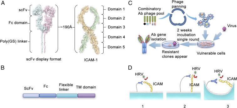
Fig. 1.
Schematic representation of selection of Rhinovirus blocking, ICAM-binding antibodies. (A) Predicted structure and dimension of ICAM-1 ectodomain and the membrane tethered antibody. (B) The construct used to express antibody on the cell surface. (C) Phage displayed followed by the functional selection to identify the HRV-blocking antibodies. (D) Three models of proposed interactions between ICAM and membrane-tethered antibodies.

