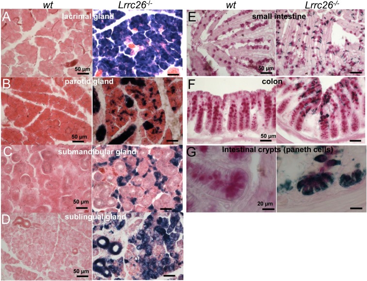
Fig. 2.
Bluo-Gal staining is observed in glandular acinar cells and goblet and Paneth cells of gastrointestinal tract. In A–D, tissues were developed for Bluo-Gal staining and counterstained with eosin. (A) LRRC26 KO tissues show abundant Bluo-Gal reaction product throughout a lacrimal gland section that is absent in wt sections. (B) In parotid, Bluo-Gal reaction product (Right) is observed sparsely, but throughout acinar cells, whereas dense staining likely corresponds to intralobular and interlobular parotid ducts. (C) Bluo-Gal staining in submandibular gland is confined to cells likely to be seromucous acinar cells, whereas larger glandular duct cells display little if any staining. (D) Bluo-Gal staining in sublingual gland is distributed throughout acinar cells and ducts within the gland. In E–G, tissues were first processed with Bluo-Gal staining followed by a periodic acid-Schiff (PAS) reaction. (E) Goblet cells in villi of the small intestine are positive for PAS staining at the apical end corresponding to mucus granules and positive for Bluo-Gal staining at the basal end. (F) Crypts in the colon exhibit abundant PAS-positive cells, with the most superficial cells also enriched with Bluo-Gal staining. (G) Crypts of the small intestine reveal positive PAS staining and Bluo-Gal staining in Paneth cells and goblet cells.

