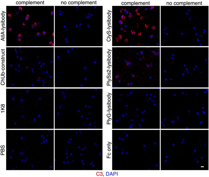
Fig. 3.
Lysibodies induce deposition of complement on the surface of S. aureus cells. S. aureus Wood 46 cells (protein A negative) were attached to poly-l-lysine–coated coverslips and incubated with lysibodies. The cells were then incubated with human complement, washed, fixed, and blocked. Complement was detected using rabbit anti-C3, followed by Alexa Fluor 594 conjugate; DNA was stained with DAPI. Images were obtained using deconvolution microscopy and are presented as maximum intensity projections. Experiments were repeated twice (scale bar, 1 μm).

