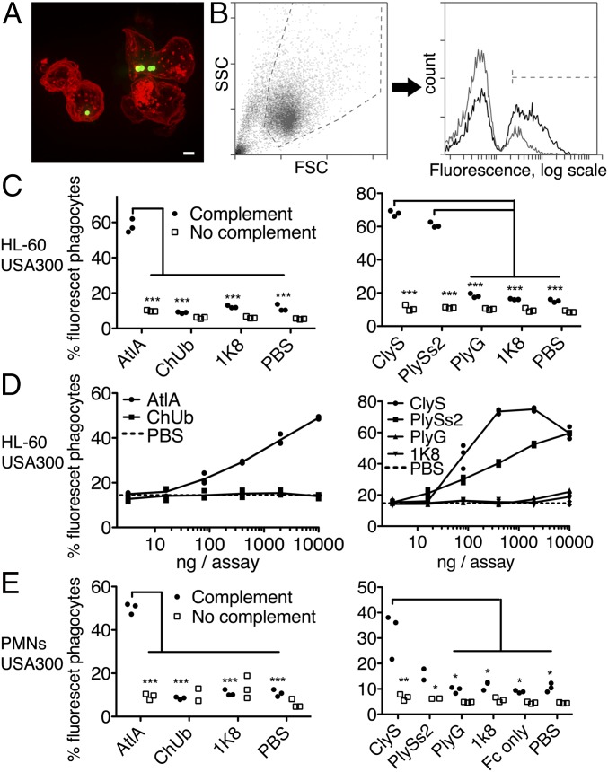
Fig. 4.
Lysibodies induce phagocytosis of S. aureus by neutrophils. HL-60 neutrophils (A–D) and human polymorphonuclear cells (E) were incubated with FITC-labeled S. aureus strain USA300 in the presence of lysibodies and S. aureus-adsorbed human complement. (A) A representative image of HL-60 neutrophils incubated with FITC-labeled S. aureus USA300 and AtlA lysibody; a maximum intensity projection is presented (scale bar, 2 μm). Also see Fig. S9. (B) Gating scheme for flow cytometry analysis: gating on neutrophils using forward and side scatter (Left) and determination of the percentage of fluorescent neutrophils (Right). Black, AtlA lysibody; gray, PBS. (C) Phagocytosis of S. aureus by HL-60 neutrophils using 5 µg AtlA lysibody, ClyS lysibody, PlySs2 lysibody, or controls in the presence or absence of complement. (D) Effect of lysibody dose on S. aureus phagocytosis by HL-60 neutrophils. (E) Phagocytosis of S. aureus by human polymorphonuclear cells using 5 µg AtlA lysibody, ClyS lysibody, PlySs2 lysibody, or controls in the presence or absence of complement. All experiments were done in triplicates and repeated two to four times. Statistical significance analysis using the t test was performed for the relevant samples. *P < 0.05, **P < 0.01, ***P < 0.001. Also see Fig. S10 (Wood 46 and USA600). FSC, forward scatter; PMNs, polymorphonuclear leukocytes; SSC, side scatter.

