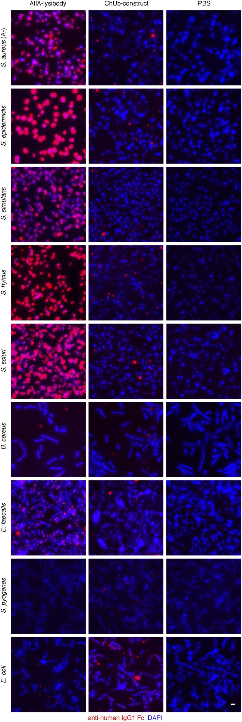
Fig. S5.
Binding range of AtlA lysibody. The following strains were evaluated for binding of lysibodies using fluorescence microscopy: S. aureus protein A negative Wood 46, S. epidermidis ATCC 12228S, S. simulans TNK3, S. hyicus HER1048, S. sciuri subsp. sciuri K1, B. cereus T, E. faecalis V12, S. pyogenes SF370, and E. coli DH5α. Bacterial cells were fixed, attached to a microscope cover glass, and blocked. Cells were incubated with AtlA lysibody or ChUb construct and subsequently with anti-human IgG Fc Alexa Fluor 594 conjugate. DNA was visualized using DAPI (scale bar, 2 μm).

