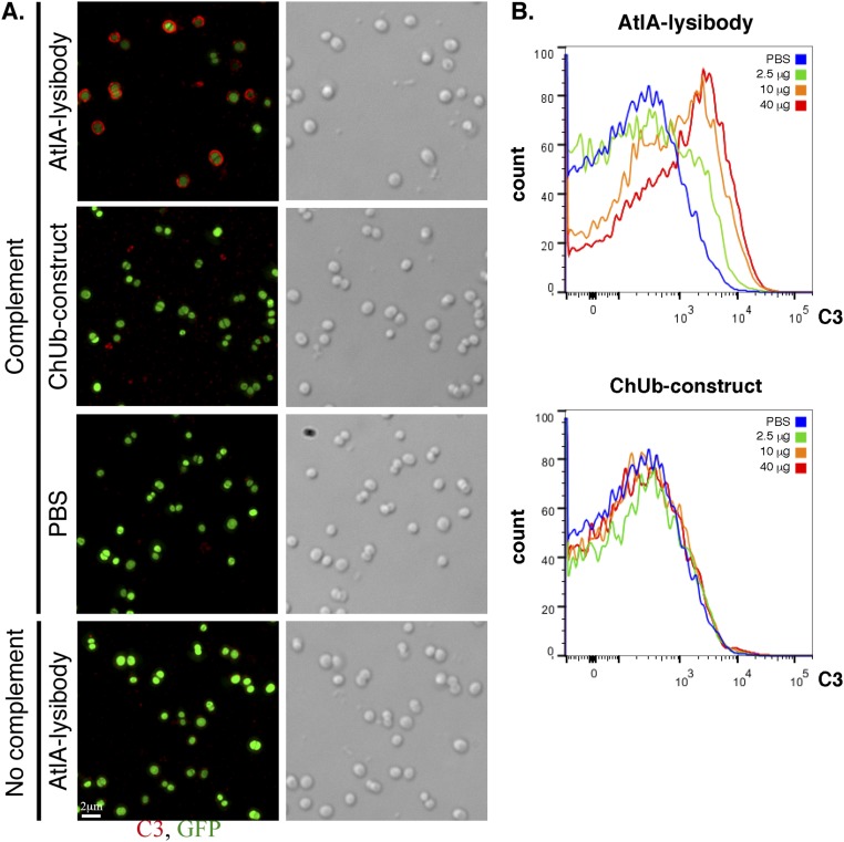
Fig. S8.
AtlA lysibody induces deposition of complement on the surface of S. aureus cells. (A) S. aureus Newman/pCN57 (GFP) cells were attached to a poly-l-lysine–coated coverslips and incubated with AtlA lysibody or ChUb construct and then with S. aureus-adsorbed human complement. The cells were washed, fixed, and blocked (protein A was blocked with heat-inactivated serum). Complement was detected using rabbit anti-C3, followed by Alexa Fluor 594 conjugate. Deconvolution microscopy images are presented as maximum intensity projections (scale bar, 2 μm). (B) S. aureus Newman/pCN57 (GFP) cells were incubated with various concentrations of AtlA lysibody or ChUb construct, washed, and incubated with S. aureus-adsorbed human complement. The cells were then washed, fixed, and blocked (protein A was blocked with heat-inactivated serum). Complement was detected using rabbit anti-C3, followed by Alexa Fluor 594 conjugate. For flow cytometry analysis, initial gating was done on GFP-expressing cells, and then the C3 fluorescence in the red channel was evaluated.

