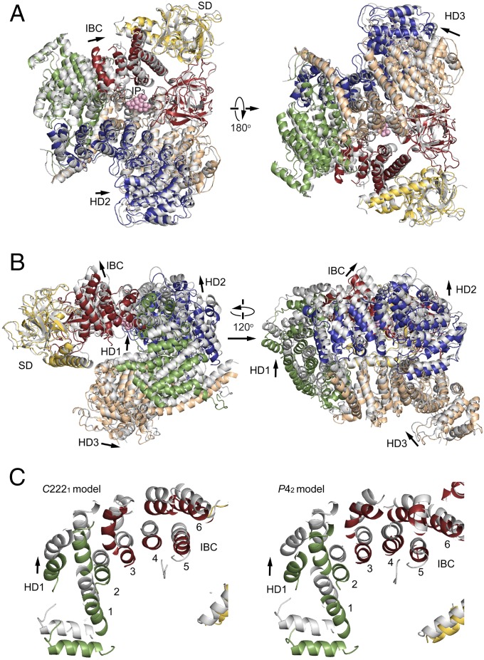
Fig. 3.
Long-range conformational changes in the large cytosolic domain by IP3. (A) Comparison of IP3R2217 structures in the absence of IP3 (colored) and in the presence of IP3 (gray). Two structures were superposed by fitting of the N-terminal β-domain (7-430 residues). (B) Side views of the two crystal structures. IP3 molecules are in pink, and arrows indicate the direction of domain relocations by IP3. (C) The helical arrangements (helices 1–6) at transitional regions from IBC to HD1 of IP3R2217 (Left) and IP3R1585 (Right) were viewed from an IP3-binding site.

