Fig. S1.
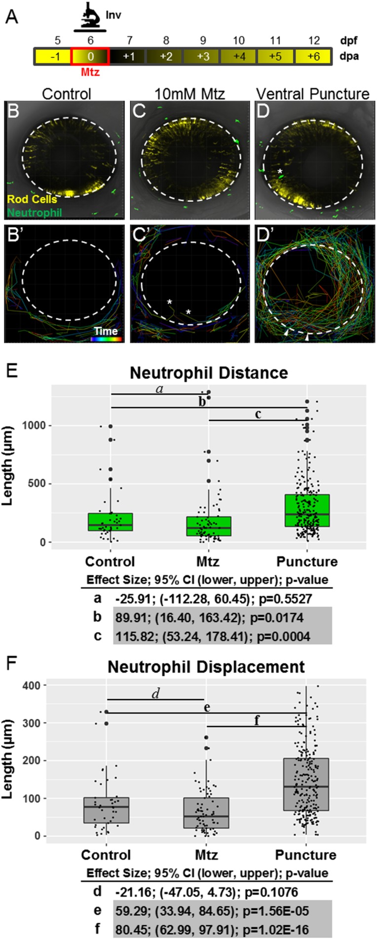
Neutrophils respond to retinal puncture wounds but not to rod cell ablation. (A) Schematic of timelines for intravital imaging assays performed in B–F. The line denotes the time during which images were collected. (B–D) Whole-retina, 20-h intravital time-lapse sequences (image interval, 20 min) were collected from 6-dpf double-transgenic rho:YFP-NTR;mpx:GFP larvae with YFP-NTR–expressing rod cells (yellow) and GFP-expressing neutrophils (green), and individual neutrophil migration paths were quantified (34× magnification). Representative z-projection confocal images of 6-dpf larvae from untreated control (B and B′), Mtz-treated (C and C′), and puncture wound (D and D′) groups. B′, C′, and D′ show the migration paths of tracked neutrophils; rainbow dragon tails show the position of each neutrophil over the 20-h time-lapse sequence (purple to red line segments). No evidence of neutrophil infiltration into the neural retina was observed in either the untreated control (B′) or rod cell-ablated (C′) condition. The asterisks mark the paths of neutrophils that migrated into extraretinal tissue over the eye. Conversely, puncture wounding (denoted by arrowheads in D′) permitted robust chemotaxis to the injury site and neutrophil infiltration into the neural retina. (E and F) Quantification of neutrophil migration patterns. Box plots show distance (E) and displacement (F) patterns across all three conditions with data from individual neutrophils displayed as a dot blot overlay (larger dots denote outliers). Increased neutrophil migration behaviors are evident only in puncture-wounded retinas. Statistical comparisons (Student’s t test) between paired groups (effect size, 95% confidence intervals, and P value) are provided in the tables below the graphs. (Note: Pairs in which CIs suggest reproducible findings are shaded gray).

