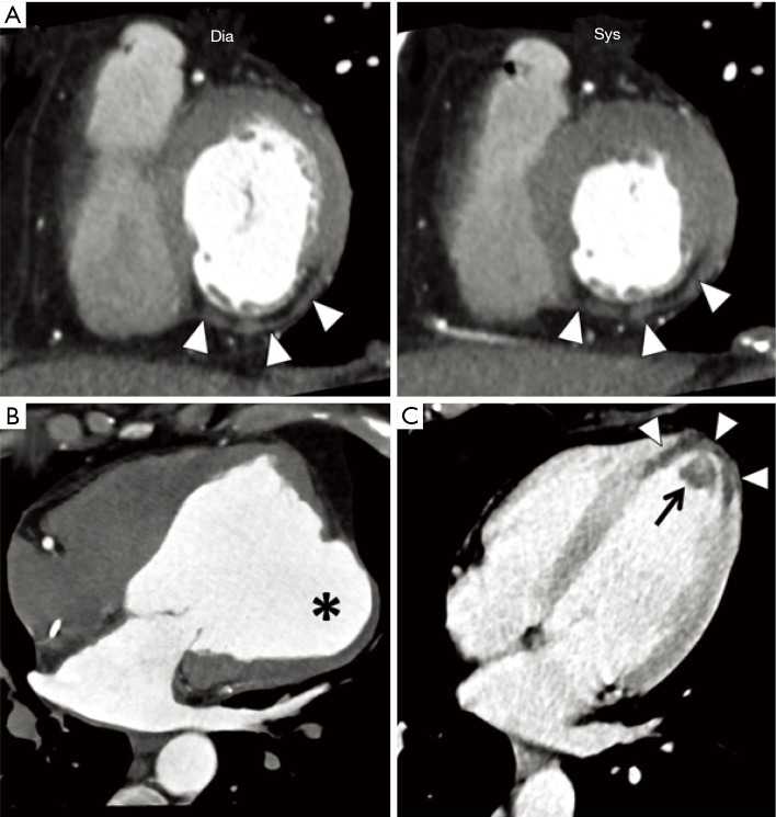
Figure 2.
CT rest perfusion imaging of myocardial infarction. (A) Myocardial wall thinning associated with sub-endocardial hypoperfusion and adipose metaplasia are known signs of chronic infarction. Lipomatous metaplasia is usually seen as hypoattenuated areas (<0 HU) mostly in the subendocardial location (arrowheads). Concomitant akinesia is indicative of transmural infarction; (B) CT coronary angiography can depict with high spatial resolution ischemic morphological alterations of the myocardium, such as chronic post-ischemic aneurysmal dilation (asterisk in B); (C) in the setting of acute myocardial infarction, CT coronary angiography may demonstrate the severely hypoperfused myocardium with transmural extension in the apex and para-apical region (arrowheads) with associated apical thrombosis (arrow). Dia, diastole; Sys, systole.

