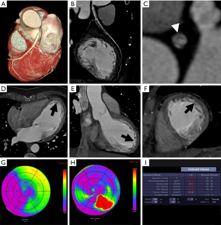
Figure 1.
CCT in a female patient with an extensive anterior myocardial infarction of the left ventricle. The patient was revascularized with a left main stenting as depicted in volume rendered (A) and multiplanar (B) images. Intra-stent intimal hyperplasia was displayed (arrowhead, C). Multiplanar images (4-chamber, D; long axis, E; short axis, F) show the first-pass perfusion defects (black arrow) on the subendocardial wall of the left ventricle. The functional bull’s eyes depict hypokinetic segments (G) and regional myocardial wall thinning (H). The left ventricular function is considerably impaired (I).

