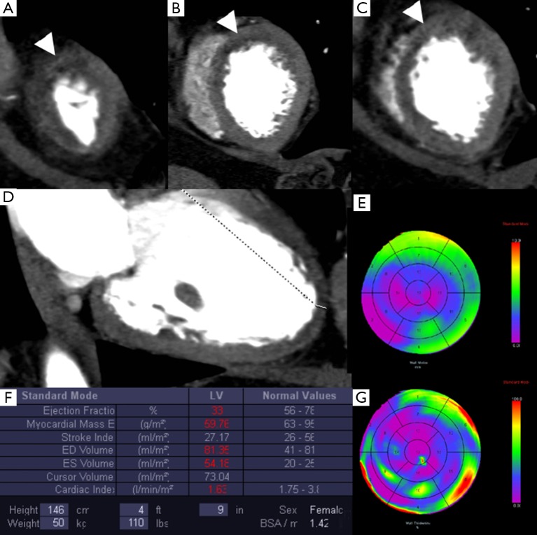
Figure 5.
Patient with a previous myocardial infarction in the territory of the left anterior descending artery. Multiplanar images in short axis (A-C) show the early perfusion defects (arrowhead) on the subendocardial wall of the left ventricle in the anterior and anteroseptal segments. In the long axis view (D) the corresponding myocardial wall is thinner than normal. The functional bull’s eyes display hypokinetic segments (E) and regional myocardial wall thinning (G). The left ventricular function is impaired (F).

