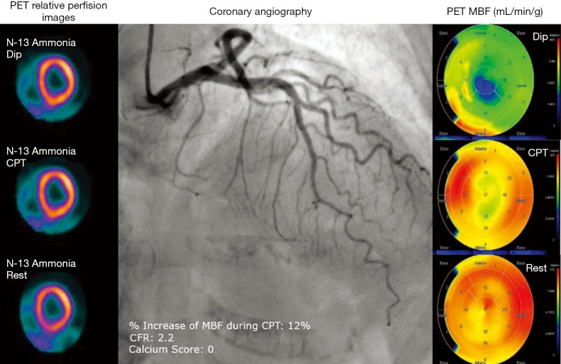
Figure 3.
A seventy-nine-year-old woman with stable angina. She has history of hypertension and hypercholesterolemia. PET short-axis cuts at rest, during CPT and during DIP induced hyperemia shows a preserved relative myocardial perfusion, in absence of coronary atherosclerosis (as shown by calcium score of 0), left panel. She exhibits a normal coronary angiogram (mid panel). Polar maps of quantification of MBF by PET shows a borderline CFR and an abnormal flow increase to CPT, suggesting that the patient has endothelial dysfunction at the level of the microcirculation (right panel). PET, positron emission tomography; CPT, cold pressor test; DIP, dipyridamole; MBF, myocardial blood flow; CFR, coronary flow reserve.

