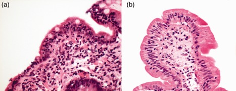
Figure 2.

(a) Close‐up of duodenal biopsy in patient with common variable immunodeficiency and chronic norovirus infection [haematoxylin and eosin (H&E) stain, ×40]. Norovirus RNA was detected by polymerase chain reaction (PCR) in stool and also from the duodenal biopsy specimen. Marked villous atrophy is apparent with a chronic inflammatory infiltrate in the lamina propria (lacking plasma cells in view of the immunodeficiency). The enterocytes are reduced in height and vacuolated. (b) Close‐up of duodenal biopsy in the same patient following norovirus clearance with a prolonged course of ribavirin (H&E stain, ×40). Note that the inflammatory infiltrate in the lamina propria has resolved, there is restitution of villous architecture and the enterocytes are columnar.
