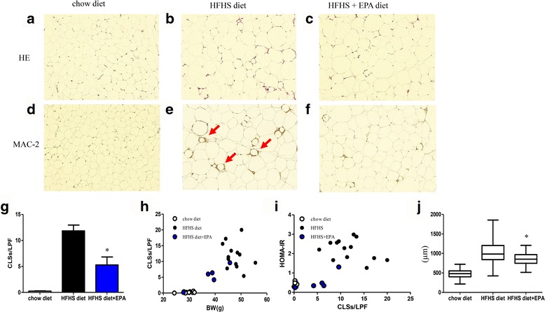
Fig. 5.

Histological analysis of mouse epididymal adipose tissue including HE staining and MAC-2 staining (a–f). Red arrows indicate CLS formation (e). CLS number per LPF in chow diet, high-fat/high-sucrose (HFHS) diet, and HFHS diet + eicosapentaenoic acid-fed mice groups (g). Correlation between body weight and CLS number (r = 0.80, P < 0.001) (h), and CLS number and HOMA-IR (r = 0.72, P < 0.001) (l). Mean adipocyte diameter in each group (j). n = 5–13 per group. Data are presented as means ± SEM. *P < 0.05 vs. HFHS diet group
