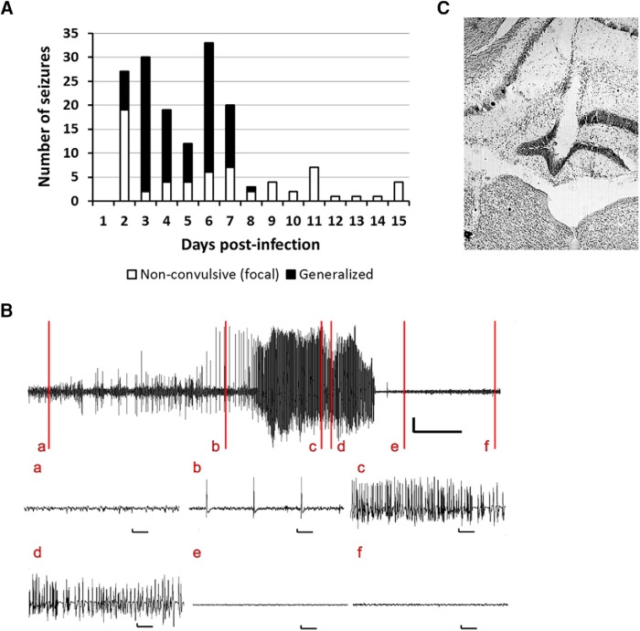
Figure 2.
Mice implanted with an electrode in the dentate gyrus for 24 h/7 d/wk vEEG show focal as well as generalized seizures after TMEV infection. (A) Total number of acute seizures each day after TMEV infection from all mice that developed acute seizures. Of 16 mice implanted with an electrode in the dentate gyrus and infected with 3 × 10⁵ pfu TMEV, 11 developed acute seizures. The mice had nonconvulsive focal seizures (white bar) as well as spontaneous generalized seizures (black bar) during the first week after TMEV infection; however, nonconvulsive focal seizures continued to occur in some mice after acute behavioral seizures subsided. Although the vEEG was conducted for more than 1 mo postinfection, the seizure data are shown until 15 dpi, as several mice were discontinued from the study after 15 dpi owing to loss of electrode assembly. (B) Representative trace of vEEG recording from a mouse at 5 d after TMEV infection. The vertical lines on the upper vEEG trace indicate baseline EEG during normal mouse behavior (a); nonconvulsive focal electrographic seizure (b); stage 3 behavioral seizure (c); stage 4 behavioral seizure (d); stage 4 behavioral seizure without electrographic activity (e); and normal mouse behavior and the electrographic activity returning to baseline (f). All vertical scale bars represent 500 µV, and the horizontal scale bars represent 30 s in the upper vEEG trace and 1 s in each of the expanded traces below. (C) Location of the electrode in the molecular layer of the dentate gyrus (near the center of the image). The coronal brain slice shown here, stained with 0.1% cresyl violet, was obtained from a TMEV-infected mouse that had seizures.

