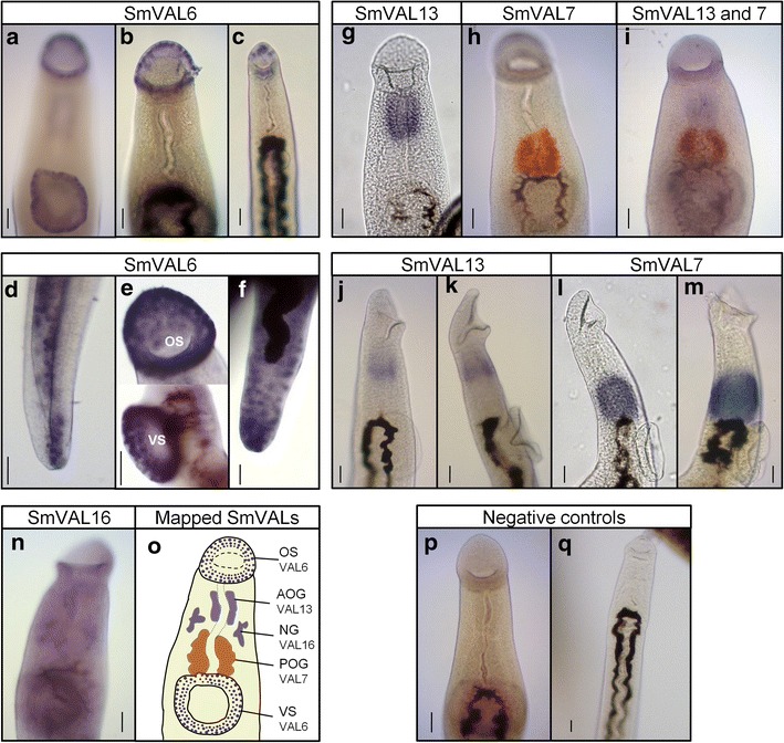
Fig. 4.

Localization of SmVAL6, 7, 13 and 16 in the anterior region of adult Schistosoma mansoni by WISH. Adult worms were analyzed using SmVAL6, 7, 13 and 16 DIG or fluorescein-labeled probes and revealed with two different substrates (BM Purple - purple, or INT/BCIP - orange). SmVAL6 transcripts identified in the oral and ventral suckers of adult male (a, b, e) and female worms (c) and in the tegument cell bodies of the posterior region of the parasite (d, f). SmVAL13 localization in the anterior oesophageal gland of 7-week-old male adult worms (g), as well as, in 3 and 5-week-old worms (j, k). SmVAL7 localization in the posterior oesophageal gland of 7-week-old male adult worms, revealed with INT/BCIP (orange) (h), as well as, in 3 and 5-week-old worms (l, m). Double in situ localization of SmVAL13 (purple) and SmVAL7 (orange) in the anterior and posterior oesophageal glands, respectively (i). SmVAL16 transcript localized close to the neural ganglia of male adult worm (n). No staining was observed in the tissues of male and female worms hybridized with the negative control (SmVAL13 sense probe) (p, q), respectively. Schematic representation of SmVALs mapped so far (o): OS, oral sucker; VS, ventral sucker; AOG, anterior oesophageal gland; POG, posterior oesophageal gland; NG, neural ganglia. Development of color was observed after approximately 4 h of the enzyme reaction for SmVAL7 and 13, 72 h for SmVAL16 and up to 12 h for SmVAL6. Scale-bars: 50 μm
