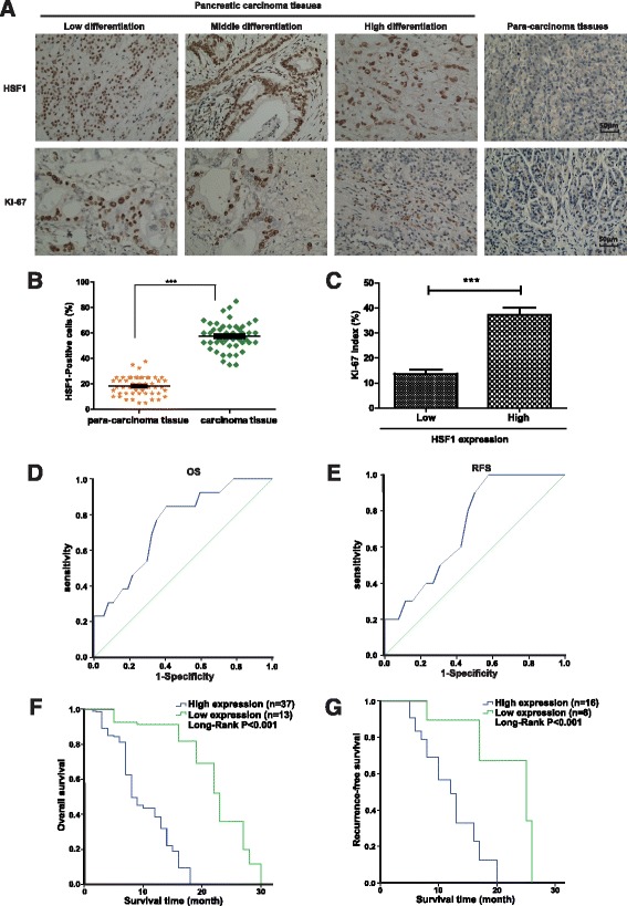
Fig. 1.

High expression of HSF1 in pancreatic cancer tissues and association with poor prognosis in patients. a Representative image of immunohistochemical detection of the expression of HSF1 and Ki-67 proteins in 50 pairs of pancreatic cancer and para-carcinoma tissue specimens. Scale bar, 50 μm. b Semi-quantitative analysis of HSF1 protein expression in pancreatic cancer and para-carcinoma tissues. Differences were analyzed by paired t-test and data represent the mean ± standard deviation of three independent experiments. ***P < 0.001. c Correlation between HSF1 and the proliferation marker, Ki-67, in pancreatic cancer tissues by immunohistochemistry. Figure shows the Ki-67 index in pancreatic cancer tissues with low or high expression of HSF1. d and e Receiver operating characteristic (ROC) curve analysis of the correlation between the expression of HSF1 protein in patients with pancreatic cancer and overall survival (d) and recurrence-free survival (e). f and g Kaplan–Meier survival curve analysis of the correlation between the expression of HSF1 protein in patients with pancreatic cancer and overall survival (f) and recurrence-free survival (g)
