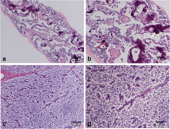
Fig. 3.

Representative hematoxylin and eosin (H&E) staining of tissue sections from CT-guided biopsy of left pulmonary a, b and left parathyroidectomy c, d. a The alveoli structure was partially damaged. Fibrosis and interalveolar septa broadening were seen in the pulmonary interstitium with multifocal calcium deposition and irregular-shaped calcified bodies. No obvious inflammatory cell or giant cell reaction was observed in pulmonary interstitium (H&E 100 × .). b Multifocal irregularities of calcium deposition and the calcified bodies in the pulmonary interstitium were seen at high magnification (white arrow), some of which resemble the psammoma bodies seen in a thyroid gland papillary carcinoma (red arrow) (H&E 200 × .). c Tumor cells were shown as the organ-like tissue structure and the tumor cells were in the nest-like distribution. Branched blood vessels were found between the cells and no tumor necrosis was observed (H&E 100 × .). d High magnification revealed the nest-like distribution of tumor cells, which were round or columnar with the cytoplasm being transparent and the nucleus being round or oval. Neither nucleus atypia nor mitotic activity was observed. Sinusoid segmentation could be found between the tumor cells (H&E 200 × .)
